Surgery
Dental Health and Wisdom Teeth
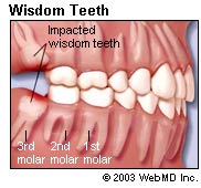
Wisdom teeth are the third and final set of molars that most people get in their late teens or early twenties. Sometimes these teeth can be a valuable asset to the mouth when healthy and properly aligned, but more often, they are misaligned and require removal.
Wisdom teeth present potential problems when they are misaligned – they can position themselves horizontally, be angled toward or away from the second molars or be angled inward or outward. Poor alignment of wisdom teeth can crowd or damage adjacent teeth, the jawbone, or nerves. Wisdom teeth that lean toward the second molars make those teeth more vulnerable to decay by entrapping plaque and debris. In addition, wisdom teeth can be entrapped completely within the soft tissue and/or the jawbone or only partially break through or erupt through the gum. Teeth that remain partially or completely entrapped within the soft tissue and /or the jawbone are termed "impacted." Wisdom teeth that only partially erupt allows for an opening for bacteria to enter around the tooth and cause an infection, which results in pain, swelling, jaw stiffness, and general illness. Partially erupted teeth are also more prone to tooth decay and gum disease because their hard-to-reach location and awkward positioning makes brushing and flossing difficult.
How Do I Know if I Have Wisdom Teeth?
Wisdom teeth present potential problems when they are misaligned – they can position themselves horizontally, be angled toward or away from the second molars or be angled inward or outward. Ask your dentist about the positioning of your wisdom teeth. He or she may take an X-ray periodically to evaluate for the presence and alignment of your wisdom teeth. Your dentist may also decide to send you to an oral surgeon for further evaluation.
Your dentist or oral surgeon may recommend that your wisdom teeth be extracted even before problems develop. This is done to avoid a more painful or more complicated extraction that might have to be done a few years later. Removal is easier in young people, when the wisdom teeth roots are not yet fully developed and the bone is less dense. In older people, recovery and healing time tend to be longer.
How Are Wisdom Teeth Removed?
 The relative ease at which your dentist or oral surgeon can extract your wisdom teeth depends on their position. Your oral health care provider will be able to give you an idea of what to expect during your pre-extraction exam. A wisdom tooth that is fully erupted through the gum can be extracted as easily as any other tooth.
However, a wisdom tooth that is underneath the gums and embedded in the jawbone will require an incision into the gums and then removal of the portion of bone that lies over the tooth. Oftentimes, for a tooth in this situation, the tooth will be extracted in small sections rather than removed in one piece to minimize the amount of bone that needs to be removed to get the tooth out.
Wisdom tooth extraction
An oral and maxillofacial surgeon or your dentist can remove (extract) a wisdom tooth. The procedure often can be done in the dentist's or surgeon's office.
If you have any infections, surgery will usually be delayed until the infection has cleared up. Your doctor or dentist may have you take antibiotics to help heal the infection.
Before removing a wisdom tooth, your dentist will give you a local anesthetic to numb the area where the tooth will be removed.
To remove the wisdom tooth, your dentist will open up the gum tissue over the tooth and take out any bone that is covering the tooth. He or she will separate the tissue connecting the tooth to the bone and then remove the tooth. Sometimes the dentist will cut the tooth into smaller pieces to make it easier to remove.
After the tooth is removed, you may need stitches. Some stitches dissolve over time and some have to be removed after a few days. Your dentist will tell you whether your stitches need to be removed. A folded cotton gauze pad placed over the wound will help stop the bleeding.
Frenectomy
A frenectomy is the surgical removal of frenulum or frenum (frenae - plural), which is a thin band of tissue found in various parts of the body, especially the mouth. A frenectomy is a common surgical procedure in dental and orthodontic practices. Children and people being fitted for dentures are the most frequent candidates for a frenectomy.
There are two primary locations in the mouth where frenum is found – under the tongue and underneath the center of the upper lip. The frenum attaches the muscles of the cheeks and lips to the mouth, but in some cases, this tissue may interfere with the development of the mouth. In the event that the tissue, or frenum, is attached to close to the tip of the tongue or too far down the gums between the front teeth, a frenectomy may be performed.
A lingual frenectomy is the removal of the lingual frenum, or the tissue under the tongue. Generally, if the tissue is attached too closely to the tip of the tongue, it can interfere with speech development and proper tooth development. A lingual frenectomy is a fairly common procedure for children who may be “tongue tied” and is sometimes referred to as clipping the tongue. After the procedure, the tongue can usually be fully extended and becomes fully mobile.
A labial frenectomy is the removal of the tissue attached to the center of the upper lip. Frenum attached too far down the gum can cause gum recession and gaps between the front teeth. Further, denture patients often have a labial frenectomy to achieve a proper denture fit.
A frenectomy is a simple procedure performed in the dentist or orthodontist’s office. Patients rarely experience any complications from the procedure and it can often correct problems that have or could occur due to excess tissue. Though a frenectomy is a common oral surgical procedure, frenum is also found in other parts of the body including external genitalia, the digestive tract, and the brain. However removal of this tissue in areas other than the mouth are not common.
Sinus Lifting
A sinus lift (a sinus augmentation) is surgery that adds bone to your upper jaw in the area of your molars and premolars to make it taller. The bone is added between your jaw and the maxillary sinuses, which are on either side of your nose. To make room for the bone, the sinus membrane has to be moved upward, or "lifted." A sinus lift usually is done by an oral and maxillofacial surgeon or a periodontist.
It can happen that there is no longer enough bone in the molar area of the maxilla in the direction of the floor of the sinus because of the bone atrophy after tooth loss. As bone augmentation in this region is possible only with difficulty, a method was developed in which the floor of the sinus is raised and bone is inserted into the cavity produced, without injuring the mucosa of the sinus, which leads to an effective increase in the bone in the molar maxillary region. This operation is called a sinus lift. This operation can be carried out under local anaesthesia and is done through the mouth so that there are no scars on the face. The sinus lift operation can often be done at the same time as the insertion of dental implants. The bone material which is inserted corresponds to that described already for bone augmentation.
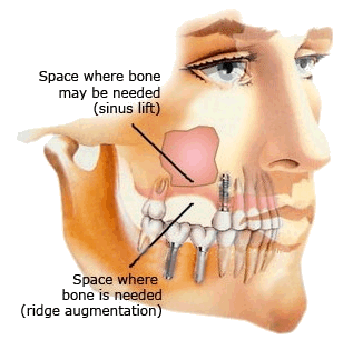
A sinus lift is done when there is not enough bone in the upper jaw, or the sinuses are too close to the jaw, for dental implants to be placed. There are several reasons for this:
- Many people who have lost teeth in their upper jaw - particularly the molars teeth - do not have enough bone for implants to be placed. Because of the anatomy of the skull, the back of the upper jaw has less bone than the lower jaw.
- Once teeth are gone, bone begins to be resorbed (absorbed back into the body). If teeth have been missing for a long time, there often is not enough bone left to place implants.
- The maxillary sinus may be too close to the upper jaw for implants to be placed. The shape and the size of this sinus varies among individuals. In addition, the sinus can get larger as you age.
- Bone may have been lost because of periodontal (gum) disease.
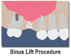 Sinus Lift Procedure The maxillary sinuses are air-filled cavities above your back teeth in the upper jaw. Following tooth loss and subsequent bone loss in that area, there is often insufficient bone height in the back of the upper jaw to place dental implants. This procedure involves elevating or "lifting" the lining of the sinus cavity and placing the bone graft onto the sinus floor. This essentially increases the amount of bone above the back of your upper jaw, allowing implants to now be placed.